Blatt Bioimaging Center
Already have a Logon Account?
|
Welcome
Welcome to Syracuse University FBS Portal. This site is designed to automate the use of our Core Facilities and to provide the best possible customer service. Quick Info
For more info, please contact the Priority Software Support Team. | Our Core Facilities To learn more about a particular facility or to request access, please click on a facility name below. |
Main Contact Info
Abrar Aljiboury
234 Life Sciences Complex
Syracuse University
Syracuse, NY 13244
Remittance Contact Info
Abrar Aljiboury
Blatt BioImaging Center
359 Life Sciences Complex
Syracuse, NY 13244
This facility has not published any Products. Please check back.
The following Products and Services are available within our facility:
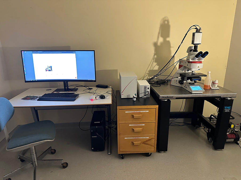
Instruments
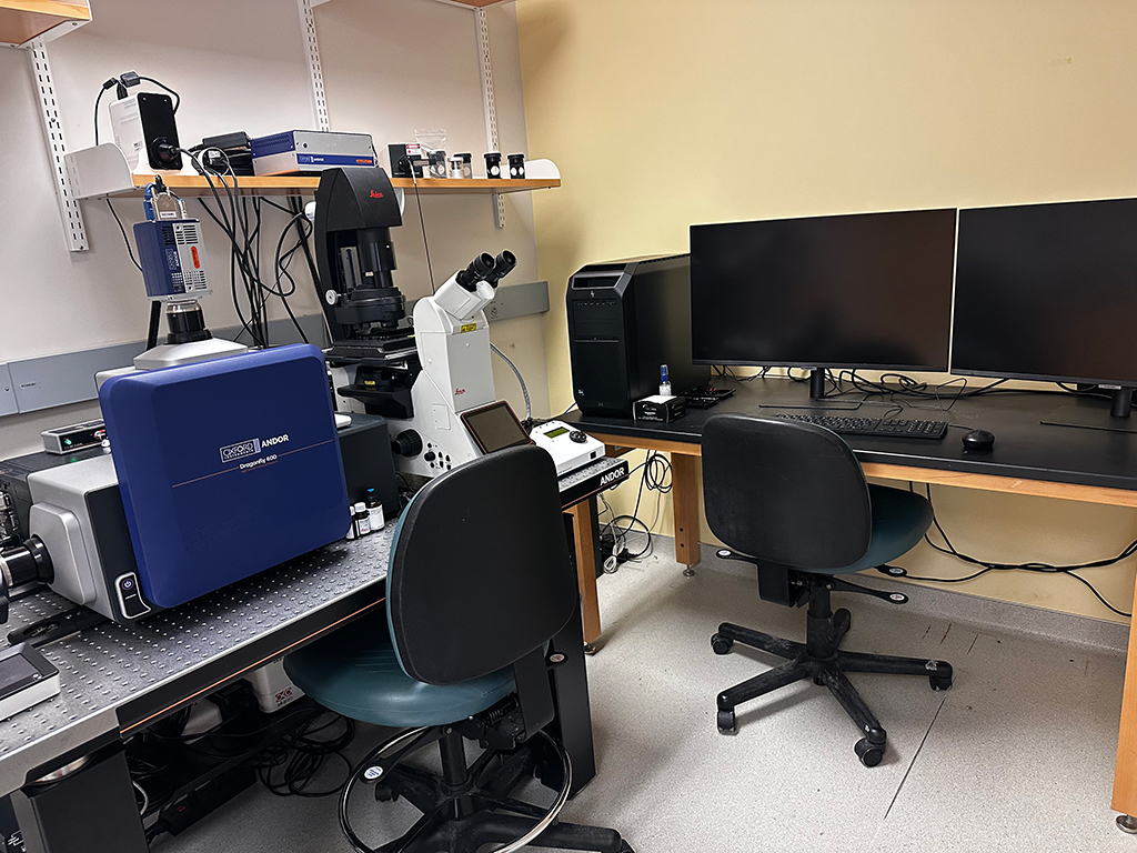 |
Dfly620-SR• Widefield and fluorescence |
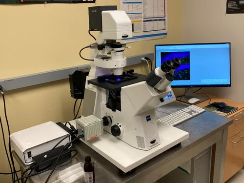 |
AXIO-OBZ1
|
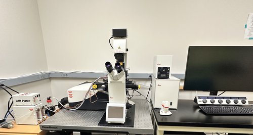 |
DMi8
|
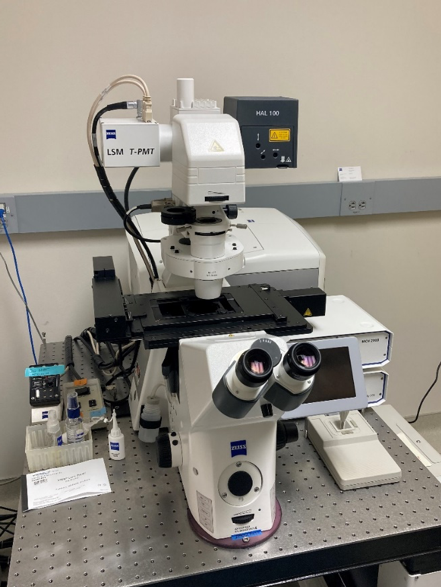 |
LSM710
|
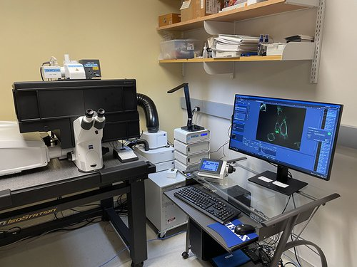 |
LSM980
|
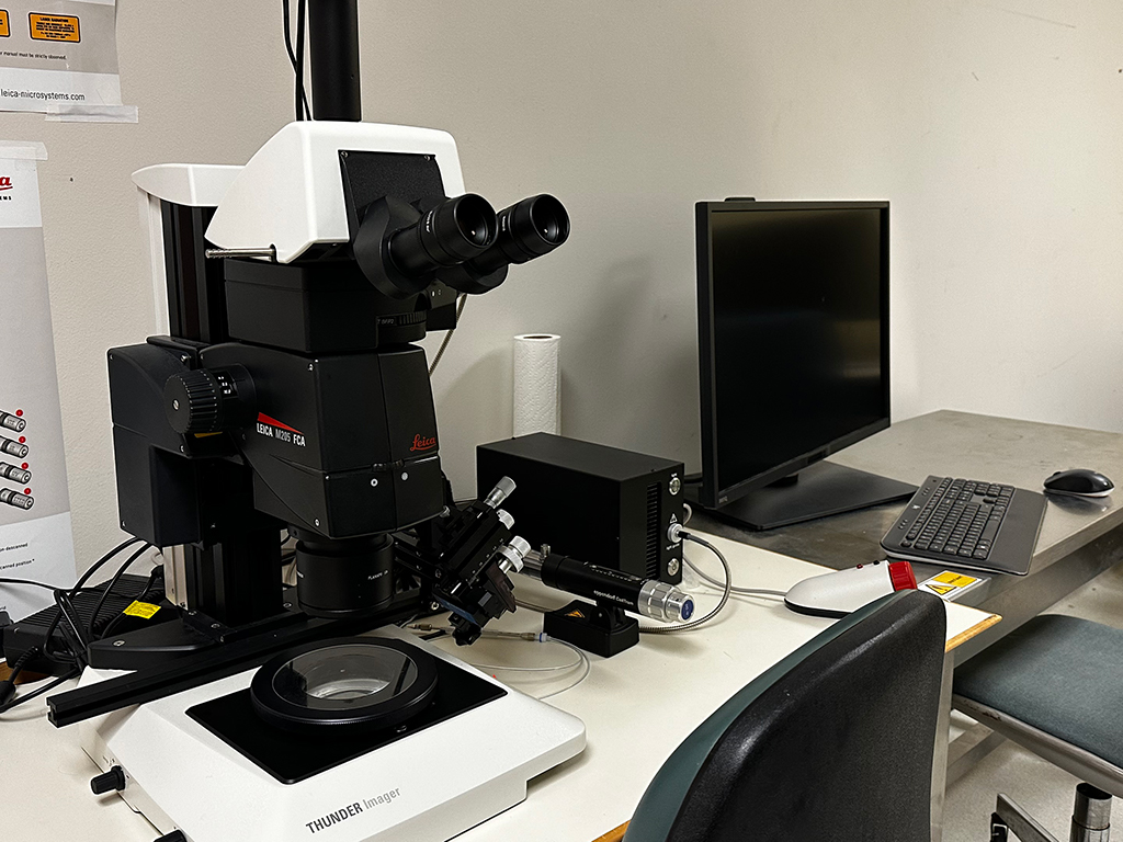 |
THUND-MOThe Leica Thunder microscope system which allows for computational clearing of thick samples such as model organisms. The Thunder system features high resolution black and white (Leica DFC9000 GT) mounted on an upright fluorescence Stereo Microscope (Leica DM6B). The system is also equipped with a micromanipulator/injection apparatus. |
 |
THUND-TThe Leica Thunder microscope system which allows for computational clearing of thick and thin samples that are stained with fluorescent markers. The Thunder system features high resolution black and white (Leica DFC9000 GT) and color cameras (Leica DMC4500) mounted on an upright fluorescence microscope (Leica DM6B). The system also features an array of objectives including long working distance, dry, water immersion, oil immersion and glycerol immersion. |
This facility has not published any News. Please check back.
Quick Quotes have not been configured. Please check back soon (Code 001, Code 002)
, please enter that email address here.")